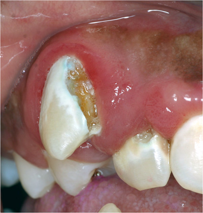
 This photo shows a good amount of decay on the facial surface of tooth #6. The chalky white area on the facial surface is demineralized enamel. The brownish colored area is demineralized dentin (cavity is within the dentin).
Tooth #7 also shows demineralization and decay on the facial surface.
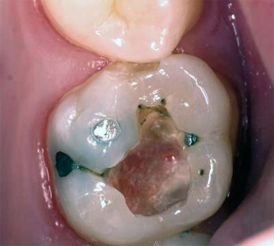
The photo to the right is showing the inside of a tooth during a restorative procedure. An occlusal amalgam was removed (due to recurrent decay) and the decay removed. The cavity was deep and extended towards the pulp of the tooth. If you look closely, you can see that the deepest portion of the preparation floor is red in color. The red color is telling you that the pulp is very close. Look even closer- do you see the small red dot that is closer to the buccal side of the tooth? That's a pulp horn that's showing.


Date last modified: May 14, 2012.
Created with SoftChalk LessonBuilder
![]()